Blog List

Thursday, 10 March 2016

CORTEX (BOTANY)

cortex is the outermost layer of a stem or root in a plant, or the surface layer or "skin" of the nonfruiting part of the body of some lichens.
In botany, the cortex is the outermost layer of the stem or root of a plant, bounded on the outside by the epidermis and on the inside by the endodermis. In plants, it is composed mostly of differentiated cells,  usually large thin-walled parenchyma cells of the ground tissue system. The outer cortical cells often acquire irregularly thickened cell walls, and are called collenchyma cells. Some of the outer cortical cells may contain chloroplasts. It is responsible for the transportation of materials into the central cylinder of the root through diffusion and may also be used for food storage in the form of starch.
Cross-section of a flax plant stem:
1. Pith
2. Protoxylem
3. Xylem I
4. Phloem I
5. Sclerenchyma (bast fibre)
6. Cortex
7. Epidermis
On a lichen the cortex is the "skin", or outer layer of thallus tissue that covers the undifferentiated cells of the medulla. Fruticose lichens have one cortex encircling the branches, even flattened, leaf-like forms; foliose lichens have different upper and lower cortices; crustose, placodioid and squamulose lichens have an upper cortex but no lower cortex; and leprose lichens lack any cortex.
References

  1. ^ What is a lichen?, Australian National Botanical Garden

- Wikipedia 

EPIDERMIS (BOTANY)

The epidermis cells (from the Greek "επίδερμίδα", meaning "over-skin") is a single-layer of cells that covers the leaves, flowers, roots and stems of plants.  It forms a boundary between the plant and the external environment. The epidermis serves several functions, it protects against water loss, regulates gas exchange, secretes metabolic compounds, and (especially in roots) absorbs water and mineral nutrients. The epidermis of most leaves shows dorsoventral anatomy: the upper (adaxial) and lower (abaxial) surfaces have somewhat different construction and may serve different functions. Woody stems and some other stem structures produce a secondary covering called the periderm that replaces the epidermis as the protective covering.


Cross-section of a flax plant stem:
1. pith
2. protoxylem
3. xylem
4. phloem
5. sclerenchyma (bast fibre)
6. cortex
7. epidermis

Description
The epidermis is the outermost cell layer of the primary plant body. In some older works the cells of the leaf epidermis have been regarded as specialized parenchyma cells, but the established modern preference has long been to classify the epidermis as dermal tissue, whereas parenchyma is classified as ground tissue. The epidermis is main component of the dermal tissue system of leaves (diagrammed below), and also stems, roots, flowers, fruits, and seeds; it is usually transparent (epidermal cells have a lower number of chloroplasts or lack them completely, except for the guard cells.)


The cells of the epidermis are structurally and functionally variable. Most plants have an epidermis that is a single cell layer thick. Some plants like Ficus elastica and Peperomia, which have periclinal cellular division within the protoderm of the leaves, have an epidermis with multiple cell layers. Epidermal cells are tightly linked to each other and provide mechanical strength and protection to the plant. The walls of the epidermal cells of the above ground parts of plants contain cutin, and are covered with a cuticle. The cuticle reduces water loss to the atmosphere, it is sometimes covered with wax in smooth sheets, granules, plates, tubes or filaments. The wax layers give some plants a whitish or bluish surface color. Surface wax acts as a moisture barrier and protects the plant from intense sunlight and wind. The underside of many leaves have a thinner cuticle than the top side, and leaves of plants from dry climates often have thickened cuticles to conserve water by reducing transpiration.
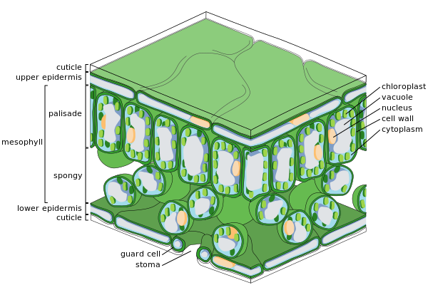
Diagram of fine scale leaf internal anatomy

The epidermal tissue includes several differentiated cell types: epidermal cells, guard cells, subsidiary cells, and epidermal hairs (trichomes). The epidermal cells are the most numerous, largest, and least specialized. These are typically more elongated in the leaves of monocots than in those of dicots.

Diagram of moderate scale leaf anatomy
Trichomes or hairs grow out from the epidermis in many species. In root epidermis, epidermal hairs, termed root hairs are common and are specialized for absorption of water and mineral nutrients.
In plants with secondary growth, the epidermis of roots and stems is usually replaced by a periderm through the action of a cork cambium.
Guards Cells
The leaf and stem epidermis is covered with pores called stomata (sing., stoma), part of a stoma complexconsisting of a pore surrounded on each side by chloroplast-containing guard cells, and two to four subsidiary cells that lack chloroplasts. The stoma complex regulates the exchange of gases and water vapor between the outside air and the interior of the leaf. Typically, the stomata are more numerous over the abaxial (lower) epidermis of the leaf than the (adaxial) upper epidermis. An exception is floating leaves where most or all stomata are on the upper surface. Vertical leaves, such as those of many grasses, often have roughly equal numbers of stomata on both surfaces. The stoma is bounded by two guard cells. The guard cells differ from the epidermal cells in the following aspects:
  • The guard cells are bean-shaped in surface view, while the epidermal cells are irregular in shape
  • The guard cells contain chloroplasts, so they can manufacture food by photosynthesis (The epidermal cells do not contain chloroplasts)
  • Guard Cells are the only epidermal cells that can make sugar. According to one theory, in sunlight the concentration of potassium ions (K+) increases in the guard cells. This, together with the sugars formed, lowers the water potential in the guard cells. As a result, water from other cells enter the guard cells by osmosis so they swell and become turgid. Because the guard cells have a thicker cellulose wall on one side of the cell, i.e. the side around the stomatal pore, the swollen guard cells become curved and pull the stomata open.

Stoma in a tomato leaf (microscope image)
At night, the sugar is used up and water leaves the guard cells, so they become flaccid and the stomatal pore closes. In this way, they reduce the amount of water vapour escaping from the leaf.
Cells Differentiation in the Epidermis

The plant epidermis consists of three main cell types: pavement cells, guard cells and their subsidiary cells that surround the stomata and trichomes, otherwise known as leaf hairs. The epidermis of petals also form a variation of trichomes called conical cells. These cells all develop from the pavement cells, which make up the majority of the plants surface cells. In short, cellular differentiation of the epidermal cells is controlled by two major factors: genetics and environmental conditions.
Trichomes develop at a distinct phase during the actual leaf development, under the control of two major trichome specification genes: TTG and GL1. The process may be controlled by the plant hormones gibberellins and even if not completely controlled, gibberellins certainly have an effect on the development of the leaf hairs. GL1 causes endoreplication, the replication of DNA without subsequent cell division as well as cell expansion. GL1 turns on the expression of a second gene for trichome formation, GL2, which controls the final stages of trichome formation causing the cellular outgrowth.
Arabidopsis thaliana uses the products of inhibitory genes to control the patterning of trichomes, such as TTG and TRY. The products of these genes will diffuse into the lateral cells, preventing them from forming trichomes and in the case of TRY promoting the formation of pavement cells.
As previously mentioned, conical cells are a form of trichome that occurs on the petals of flowers . Expression of the gene MIXTA, or its analogue in other species, later in the process of cellular differentiation will cause the formation of conical cells over trichomes. MIXTA is a transcription factor.
Stomatal patterning is a much more controlled process, as the stoma effect the plants water retention and respiration capabilities. As a consequence of these important functions, differentiation of cells to form stomata is also subject to environmental conditions to a much greater degree than other epidermal cell types.
Stomata are holes in the plant epidermis that are surrounded by two guard cells, which control the opening and closing of the aperture. These guard cells are in turn surrounded by subsidiary cells which provide a supporting role for the guard cells.
Stomata begin as stomatal meristemoids. The process varies between dicots and monocots. Spacing is thought to be essentially random in dicots though mutants do show it is under some form of genetic control, but it is more controlled in monocots, where stomata arise from specific asymmetric divisions of protodermal cells. The smaller of the two cells produced becomes the guard mother cells. Adjacent epidermal cells will also divide asymmetrically to form the subsidiary cells.
Because stomata play such an important role in the plants survival, collecting information on their differentiation is difficult by the traditional means of genetic manipulation, as stomatal mutants tend to be unable to survive. Thus the control of the process is not well understood. Some genes have been identified. TMM is thought to control the timing of stomatal initiation specification and FLP is thought to be involved in preventing further division of the guard cells once they are formed.
Environmental conditions affect the development of stomata, in particular their density on the leaf surface. It is thought that plant hormones, such as ethylene and cytokines, control the stomata’s developmental response to the environmental conditions. Accumulation of these hormones appears to cause increased stomatal density such as when the plants are kept in closed environments.
References

  1. ^ Hill, J. Ben; Overholts, Lee O; Popp, Henry W. Grove Jr., Alvin R. Botany. A textbook for colleges. Publisher: MacGraw-Hill 1960
  2. ^ Evert, Ray F; Eichhorn, Susan E. Esau's Plant Anatomy: Meristems, Cells, and Tissues of the Plant Body: Their Structure, Function, and Development. Publisher: Wiley-Liss 2006. ISBN 978-0471738435
  3. ^ Raven, Peter H.; Evert, Ray F.; Curtis, Helena (1981), Biology of plants, New York, N.Y.: Worth Publishers, pp. 427–28, ISBN 0-87901-132-7, OCLC 222047616

- Wikipedia 

PARENCHYMA

Parenchyma is the bulk of a substance. In animals, a parenchyma comprises the functional parts of an organ and in plants parenchyma is the ground tissue of nonwoody structures.
The term parenchyma is New Latin, f. Greek παρέγχυμα - parenkhuma, "visceral flesh", f. παρεγχεῖν - parenkhein, "to pour in" f. para-, "beside" + en-, "in" + khein, "to pour".
In Plants

In plants, "parenchyma" is one of the three main types of ground tissue, and the most common. It can be distinguished through their thin cell wall as compared to other cells. Parenchyma cells make up the bulk of the soft parts of plants, including the insides of leaves, flowers and fruits (but not the epidermis or veins of these structures).


Parenchyma (pale grey) in a plant stem, with scattered veins (darker red

In Animals
The parenchyma are the functional parts of an organ in the body. This is in contrast to the stroma, which refers to the structural tissue of organs, namely, the connective tissues.
In the brain, the parenchyma refers to the functional tissue in the brain that is made up of the two types of brain cell, neurons and glial cells. Damage or trauma to the brain parenchyma often results in a loss of cognitive ability or even death.
Lung parenchyma is the substance of the lung outside of the circulation system that is involved with gas exchange and includes the alveoli and respiratory bronchioles.
In cancer, the parenchyma refers to "The portion of a tissue that lies outside the circulatory system and is often responsible for carrying out the specialized functions of the tissue"
References

  1. ^ LeMone, Priscilla; Burke, Karen; Dwyer, Trudy; Levett-Jones, Tracy; Moxham, Lorna; Reid-Searl, Kerry; Berry, Kamaree; Carville, Keryln; Hales, Majella; Knox, Nicole; Luxford, Yoni; Raymond, Debra (2013). "Parenchyma".  Medical-Surgical Nursing. Pearson Australia. p. G–18. ISBN 978-1-4860-1440-8.
  2. ^ "Parenchyma". Retrieved 30 December 2014.
  3. ^ http://www.wisegeek.org/what-is-the-brain-parenchyma.html.
  4. ^ "Lung parenchyma. Retrieved 9 February 2016.
  5. ^ Weinberg (c. 2014). The Biology of Cancer.


- Wikipedia 

Pineapple Juice & Pregnancy

Pineapple juice can be a healthy addition to a pregnancy diet, especially since it contains vital nutrients that your developing baby needs to remain healthy. While some pregnant women worry about the safety of pineapple juice during pregnancy, these fears are mostly based on misconceptions about the effect of the pineapple component bromelain on the cervix.
Pineapple Juice & Pregnancy
Pineapple juice is a healthy beverage during pregnancy. Photo Credit HandmadePictures/iStock/Getty Images

Pineapple Juice

Pineapple juice is extracted from fresh pineapples, a fruit native to South America. Pineapple is high in vitamin C, also known as ascorbic acid. Vitamin C is an essential nutrient that helps build bones, skin, cartilage and tendons in your developing baby. Pregnant women need at least 85mg of vitamin C each day, and pineapple juice contains 25mg per cup. Pineapple juice also provides plenty of other nutrients, including vitamins A and B-6, folate, niacin, thiamin, riboflavin, pantothenic acid, iron, magnesium, potassium and manganese. Fresh pineapple might be healthier than pineapple juice, however, since it also contains dietary fiber.

Safety

Pineapple was traditionally used to induce miscarriage in early pregnancy, but there is no evidence that drinking pineapple juice causes pregnancy loss. Drinking pineapple juice provides plenty of beneficial nutrients and calories you need to help your baby grow, so it is generally considered safe and healthy for a pregnancy diet when consumed in moderate amounts. The acidity of pineapple juice could cause heartburn in some pregnant women. According to Drugs.com, if you consume fresh juice from an unripe pineapple it may have a severe laxative effect.

Pineapple and Labor

Some old wives tales recommend consuming pineapple or pineapple juice as a way to induce labor in the last month of pregnancy. The reasoning behind this claim is that pineapple contains a compound called bromelain, which could stimulate the cervix to dilate and thin out. However, the amounts of bromelain found in fresh pineapple are too small to have a noticeable effect on the cervix and much of the bromelain content of pineapples is lost during juicing anyway.

Gestational Diabetes

Women with gestational diabetes, a form of high blood sugar that develops during pregnancy, might need to be careful about their consumption of pineapple or other juices. Fruit juices are a more concentrated source of natural sugars than whole fruits and could cause blood sugar to spike if consumed alone in large amounts. If you have gestational diabetes, you can still drink pineapple juice as a small part of a meal containing protein or fiber, since these mitigate blood sugar response. Talk to your doctor about how to include pineapple juice or other juices into your diet if you are on a gestational diabetes pregnancy diet.
www.livestrong.com

Does Pineapple Juice Help the Lining of the Uterus?

If you're having trouble getting pregnant, you're willing to try just about anything to increase your chances of success. A rumor that pops up frequently on fertility forums states that drinking pineapple juice can increase the uterine lining thickness and improve chances of implantation. Not only is this unproven, but the core of the pineapple contains ingredients that are potentially harmful during pregnancy. Drinking pineapple juice will not improve the uterine lining, since it contains no more nutrients beneficial to uterine thickness than any other food.
Does Pineapple Juice Help the Lining of the Uterus?
Pineapple juice will not increase the chance of a pregnancy test. Photo Credit Brand X Pictures/Brand X Pictures/Getty Images

Bromelain

The core of the pineapple contains bromelain, a mixture of enzymes that digests proteins. Bromelain acts as a blood thinner and may have anti-inflammatory effects. While fertility specialists sometimes give blood thinners such as baby aspirin to help with implantation, the dose is controlled. Bromelain in large doses can cause uterine contractions and interfere with implantation of an embryo, although studies on this effect also are lacking. Some cultures use bromelain to start labor in pregnancy. Pineapple juice contains little or no bromelain, which is concentrated in the pineapple's core. Do not take bromelain supplements unless your doctor recommends it.

Selenium

Several minerals in pineapple, including selenium, supposedly contribute to improving the uterine lining. A cup of pineapple juice contains just 0.2 micrograms of selenium, a tiny percentage of the 55 micrograms required for adults over age 19. The food highest in selenium, Brazil nuts, contains 544 micrograms per ounce. Before you switch your food allegiance to Brazil nuts to help you get pregnant, consider that more than 400 micrograms of selenium per day can cause selenosis, which can lead to mild nerve damage, fatigue and irritability. Most adults in the United States gets an adequate amount of selenium in their diet; in countries where the soil contains little selenium, deficiencies can develop.

Uterine Lining and Pregnancy

The uterine lining does play a part in pregnancy, even if pineapple juice doesn't improve it. You have your best chance of getting pregnant when the uterine lining reaches not only a certain thickness but also has a certain pattern. The pattern changes during the menstrual cycle; a good pattern in the first half of the cycle, before ovulation, is not a good pattern after ovulation. A trilaminar lining, also called a triple lining or a TL pattern before ovulation is best for conception. Once your body produces progesterone, your lining should change to a homogenous pattern, sometimes called an HH pattern. A lining of between 7 and 12 millimeters is optimal for conception, but people can and do get pregnant with linings as thin as 3 millimeters, Dr. Timothy Hickman of Houston IVF reports on the Fertility Today website.

Considerations

When trying to get pregnant, it's best not to focus on one aspect to the exclusion of others. An adequate uterine lining is important, but many other factors also contribute to pregnancy. Ingredients in pineapple can have both potentially helpful and harmful effects on pregnancy. No clinical studies have proved that pineapple juice has any benefit on the uterine lining. If pineapple had any benefit, fertility doctors would include it as part of their treatment regimen. Talk with your doctor about ways to improve your uterine lining and about the potential effects of pineapple.
www.livestrong.com

Dried Vegetable Chips Nutrition

Snacking is an important part of your meal plan. It helps you control hunger and can help you meet your nutrient needs. Vegetables make a healthy snack choice because they are low in calories and high in fiber. However, many dried vegetable chips may not be as healthy a snack choice as its source vegetable, but it would be better than a traditional potato chip.
Dried Vegetable Chips Nutrition
baked vegetable chips Photo Credit Laura Clay-Ballard/iStock/Getty Images.

Ingredients

There are a number of different brands of dried vegetable chips, all with different ingredients. Nuts Online features a bulk dried vegetable chip that includes sweet potatoes, squash, carrots, green beans, taro and blue sweet potato. The chips are fried in non-hydrogenated canola oil and include added dextrin and salt. Dextrin is a type of carbohydrate that helps adhere the salt to the chip.

Serving Size and Calories

Serving sizes are standardized to help you compare similar products, according to the Food and Drug Administration. One ounce is a typical serving size for chips. A 1-ounce serving of vegetable chips contains approximately 150 calories. In comparison, the same serving of Lay's potato chips also contains 150 calories, but they are higher in fat content.

Fat

One serving of dried vegetable chips contains 6 grams of total fat and 1 gram of saturated fat. The American Heart Association recommends you limit your intake of saturated fat to less than 7 percent of total calories, or less than 15.5 grams on a 2,000-calorie diet. Because the vegetable chips are made with non-hydrogenated canola oil, they have an overall low saturated fat content, which is 1 gram.

Carbohydrates and Protein

Each 1 ounce serving of dried vegetable chips contains 17 grams of carbohydrates, 3 grams of fiber, 5 grams of sugar and 1 gram of protein. Including more fiber in your diet can help improve digestion and satisfy hunger. Healthy women need 21 to 25 grams of fiber per day and healthy men need 30 to 38 grams of fiber per day. Fiber also helps regulate your digestive system, lower your cholesterol, control blood sugar and prevent weight gain, according to the American Diabetes Association.

Sodium

Each 1 ounce serving of dried vegetable chips contains 75 grams of sodium. In comparison, a 1 ounce serving of regular Lay's potato chips contains 180 milligrams of sodium. High intakes of sodium causes your body to retain fluids, increasing your blood pressure and your risk of heart attack and stroke. The U.S. Department of Agriculture recommends you limit your intake of sodium to 1,500 to 2,300 mg a day.
www.livestrong.com

The Calories in a Margarita Drink

Served over ice in a salt-rimmed glass with a wide brim, a margarita is a Mexican beverage that can help you wash down your nachos, tacos or burritos. This drink, which has ingredients that include tequila and lime juice, is characterized by its green, white or yellow color. The caloric content of a margarita is higher than that of most other cocktails.

The Calories in a Margarita Drink
A close-up of a margarita horizontal. Photo Credit Danny Hooks/iStock/Getty Images

A High Caloric Content

A 4-ounce margarita contains about 168 calories, according to the National Institute on Alcohol Abuse and Alcoholism. The institute's Rethinking Drinking website reports that, on average, margaritas contain more calories per serving size than martinis, cosmopolitans, daiquiris and mojitos. Among standard cocktails, only the pina colada -- at 490 calories per 9 ounces -- has a higher caloric content than the margarita.
www.livestrong.com

TYLOSIS (BOTANY)

tylosis (plural: tyloses) is a bladder-like distension of a parenchyma cell into the lumen of adjacent vessels in wood. Less certainly, the term tylosis is in use to summarise the physiological process and the resulting occlusion in the xylem of woody plants as response to injury or as protection from decay in heartwood. It is a key process in wall one of the Compartmentalization Of Decay In Trees (CODIT) and other woody plants.


Section of Carpinus betulus wood showing the distinctive dark areas created as a result of tylosis in response to fungal infection

Anatomy

Observed in section under a microscope, tyloses appear as balloon-like protrusions emanating from axial paratracheal parenchyma cells into xylem vessels through pits linking the two. In some types, there may be a distinct barrier between the tyloses emanating from the pits into the vessels, while they may be barely distinguishable in other cases. 

Role in Compartmentalization

Tylosis in narrow vesseled plant vessels counteracts the axial spread of fungal hyphae and other pathogens by slowing down their vertical spread with a physical barrier. A similar process occurs in gymnosperms, which block access to tracheids by closing the pits that join them to each other.
The blocked vessels also provide a defense against the radial spread of pathogens, limiting their horizontal spread through the plant stem. Protection is stronger at the boundaries where annual rings meet.
The effectiveness of both vertical and horizontal barriers is affected by the speed at which they are established by tyloses, being typically faster in healthier plants.
Role in Heartwood Reformation

As a tree grows, its cambium adds an annual increment (or ring) of new wood, and in many species of trees the older wood towards the centre of the tree becomes less important for physiological processes like water and nutrient transportation, and this is converted by the tree to heartwood. This heartwood has no active defenses against infection, but is protected from infection and decay by the blockage of the xylem vessels with tyloses and various substances such as gums, resins and waxes containing high concentrations of volatile organic compounds such as terpenes that are toxic to insect larvae and tree pathogens such as bacteria and fungi. These products are produced by the cambium and transported to the centre of the stem by cellular structures called medullary rays radiating from the center of the stem and then enter living axial paratracheal parenchyma cells. As the wood ages, the contents of the parenchyma cell burst into the dead vessel through the pit linking the two. The parenchyma cell then dies as its contents are disgorged into the empty space of the dry vessel and highly effective decay-inhibiting substances, notably tannin, are formed and absorbed by the adjacent vessel walls.

External Links

  • Pictures of tylosis in Ulmus (Elm), with detailed description of the process

Sources

  1. ^ "MSN online dictionary. Retrieved 19 February 2010.
  2. ^ Shigo, Alex L (1991). Modern Arboriculture- A systems approach to the care of trees and their associates (third (2003) ed.). pp. 56–57. ISBN 0943563097.
  3. a b Weber, K.; Mattheck, C. (2003). Manual of wood decays in trees. Romsey, Hampshire: The Arboricultural Association. pp. 27–28. ISBN 0-900978-35-X.
  4. ^ Page, Jake; Editors of Time-Life Books (1983). Forest. Planet Earth. Time-Life Books, Amsterdam. p. 176. ISBN 0 7054 0752 7

- Wikipedia 

GROUND TISSUE

The ground tissue of plants includes all tissues that are neither dermal nor vascular. It can be divided into three classes based on the nature of the cell walls. Parenchyma cells have thin primary walls and usually remain alive after they become mature. Parenchyma forms the "filler" tissue in the soft parts of plants. Collenchyma cells have thin primary walls with some areas of secondary thickening. Collenchyma provides extra structural support, particularly in regions of new growth. Sclerenchyma cells have thick lignified secondary walls and often die when mature. Sclerenchyma provides the main structural support to a plant.


Cross-section of a flax plant stem:
1. Pith
2. Protoxylem
3. Xylem I,
4. Phloem I
5. Sclerenchyma (bast fibre).
6. Cortex
7. Epidermis

Parenchyma

Parenchyma (/pəˈrɛŋkmə/, para meaning "beside", chyma meaning "in filling") is a versatile ground tissue that generally constitutes the "filler" tissue in soft parts of plants. It forms, among other things, the cortex and pith of stems, the cortex of roots, the mesophyll of leaves, the pulp of fruits, and the endosperm of seeds. Parenchyma cells are living cells and may remain meristematic at maturity—meaning that they are capable of cell division if stimulated. They have thin but flexible cellulose cell walls, and are generally polyhedral when close-packed, but can be roughly spherical when isolated from their neighbours. They have large central vacuoles, which allow the cells to store and regulate ions, waste products, and water. Tissue specialised for food storage is commonly formed of parenchyma cells.

Cross section of a leaf showing various ground tissue types
Parenchyma cells have a variety of functions:
  • In leaves, they form the mesophyll and are responsible for photosynthesis and the exchange of gases, parenchyma cells in the mesophyll of leaves are specialised parenchyma cells called chlorenchyma cells (parenchyma cells with chloroplasts).
  • Storage of starch, protein, fats, oils and water in roots, tubers (e.g. potatoes), seed endosperm (e.g. cereals) and cotyledons (e.g. pulses and peanuts)
  • Secretion (e.g. the parenchyma cells lining the inside of resin ducts)
  • Wound repair and the potential for renewed meristematic activity
  • Other specialised functions such as aeration (aerenchyma)  and support
The shape of parenchyma cells varies with their function. In the spongy mesophyll of a leaf, parenchyma cells range from near-spherical and loosely arranged with large intercellular spaces, to branched or stellate mutually interconnected with their neighbours at the ends of their arms to form a three-dimensional network, like in the red kidney bean Phaseolus vulgaris and other mesophytes. These cells, along with the epidermal guard cells of the stoma, form a system of air spaces and chambers that regulate the exchange of gases. In some works the cells of the leaf epidermis are regarded as specialised parenchymal cells, but the modern preference has long been to classify the epidermis as plant dermal tissue, and parenchyma as ground tissue.
Collenchyma

The first use of "collenchyma" (/kəˈlɛŋkməkɒ-/) was by Link (1837) who used it to describe the sticky substance on Bletia (Orchidaceae) pollen. Complaining about Link's excessive nomenclature, Schleiden (1839) stated mockingly that the term "collenchyma" could have more easily been used to describe elongated sub-epidermal cells with unevenly thickened cell walls.
Collenchyma tissue is composed of elongated cells with irregularly thickened walls. They provide structural support, particularly in growing shoots and leaves. Collenchyma tissue makes up things such as the resilient strands in stalks of celery. Collenchyma cells are usually living, and have only a thick primary cell wall made up of cellulose and pectin. Cell wall thickness is strongly affected by mechanical stress upon the plant. The walls of collenchyma in shaken plants (to mimic the effects of wind etc.), may be 40–100% thicker than those not shaken.
Cross section of collenchyma cells
There are four main types of collenchyma:
  • Angular collenchyma (thickened at intercellular contact points)
  • Tangential collenchyma (cells arranged into ordered rows and thickened at the tangential face of the cell wall)
  • Annular collenchyma (uniformly thickened cell walls)
  • Lacunar collenchyma (collenchyma with intercellular spaces)
Collenchyma cells are most often found adjacent to outer growing tissues such as the vascular cambium and are known for increasing structural support and integrity.
Collenchyma cell are very conspicuous under the microscope due to the higher refractive index of their thickened cell wall.

Sclerenchyma
Sclerenchyma is the supporting tissue in plants. Two types of sclerenchyma cells exist: fibres and sclereids. Their cell walls consist of cellulose, hemicellulose and lignin. Sclerenchyma cells are the principal supporting cells in plant tissues that have ceased elongation. Sclerenchyma fibres are of great economic importance, since they constitute the source material for many fabrics (e.g. flax, hemp, jute, and ramie).
Unlike the collenchyma, mature sclerenchyma is composed of dead cells with extremely thick cell walls (secondary walls) that make up to 90% of the whole cell volume. The term sclerenchyma is derived from the Greek σκληρός (sklērós), meaning "hard." It is the hard, thick walls that make sclerenchyma cells important strengthening and supporting elements in plant parts that have ceased elongation. The difference between fibres and sclereids is not always clear: transitions do exist, sometimes even within the same plant.
Fibres

Fibers or bast are generally long, slender, so-called prosenchymatous cells, usually occurring in strands or bundles. Such bundles or the totality of a stem's bundles are colloquially called fibres. Their high load-bearing capacity and the ease with which they can be processed has since antiquity made them the source material for a number of things, like ropes, fabrics and mattresses. The fibres of flax (Linum usitatissimum) have been known in Europe and Egypt for more than 3,000 years, those of hemp (Cannabis sativa) in China for just as long. These fibres, and those of jute (Corchorus capsularis) and ramie (Boehmeria nivea, a nettle), are extremely soft and elastic and are especially well suited for the processing to textiles. Their principal cell wall material is cellulose.


Cross section of sclerenchyma fibres
Contrasting are hard fibres that are mostly found in monocots. Typical examples are the fibres of many grasses, agaves (sisal: Agave sisalana), lilies (Yucca or Phormium tenax), Musa textilis and others. Their cell walls contain, besides cellulose, a high proportion of lignin. The load-bearing capacity of Phormium tenax is as high as 20–25 kg/mm², the same as that of good steel wire (25 kg/ mm²), but the fibre tears as soon as too great a strain is placed upon it, while the wire distorts and does not tear before a strain of 80 kg/mm². The thickening of a cell wall has been studied in Linum.  Starting at the centre of the fibre, the thickening layers of the secondary wall are deposited one after the other. Growth at both tips of the cell leads to simultaneous elongation. During development the layers of secondary material seem like tubes, of which the outer one is always longer and older than the next. After completion of growth, the missing parts are supplemented, so that the wall is evenly thickened up to the tips of the fibres.
Fibres usually originate from meristematic tissues. Cambium and procambium are their main centres of production. They are usually associated with the xylem and phloem of the vascular bundles. The fibres of the xylem are always lignified, while those of the phloem are cellulosic. Reliable evidence for the fibre cells' evolutionary origin from tracheids exists. During evolution the strength of the tracheid cell walls was enhanced, the ability to conduct water was lost and the size of the pits was reduced. Fibres that do not belong to the xylem are bast (outside the ring of cambium) and such fibres that are arranged in characteristic patterns at different sites of the shoot.
Sclereids

Sclereids are a reduced form of sclerenchyma cells with highly thickened, lignified walls. These have a shape of a star.


Fresh mount of a sclereid
They are small bundles of sclerenchyma tissue in plants that form durable layers, such as the cores of apples and the gritty texture of pears (Pyrus communis). Sclereids are variable in shape. The cells can be isodiametric, prosenchymatic, forked or elaborately branched. They can be grouped into bundles, can form complete tubes located at the periphery or can occur as single cells or small groups of cells within parenchyma tissues. But compared with most fibres, sclereids are relatively short. Characteristic examples are brachysclereids or the stone cells (called stone cells because of their hardness) of pears and quinces (Cydonia oblonga) and those of the shoot of the wax plant (Hoya carnosa). The cell walls fill nearly all the cell's volume. A layering of the walls and the existence of branched pits is clearly visible. Branched pits such as these are called ramiform pits. The shell of many seeds like those of nuts as well as the stones of drupes like cherries and plums are made up from sclereids.
Long, tapered sclereids supporting a leaf edge in Dionysia kossinskyi
These structures are used to protect other cells.
References

  1. ^ Mauseth 2012,  pp. 98–103.
  2. ^ "Parenchyma". Merriam-Webster Dictionary. Retrieved 2016-01-21.
  3. ^ "Parenchyma . OxfordDictionaries.com. OUP. Retrieved 2016-01-21.
  4. a b Leaves.
  5. ^ Jeffree CE, Read N, Smith JAC and Dale JE (1987). Water droplets and ice deposits in leaf intercellular spaces: redistribution of water during cryofixation for scanning electron microscopy. Planta 172, 20-37
  6. ^ Hill, J. Ben; Overholts, Lee O; Popp, Henry W. Grove Jr., Alvin R. Botany. A textbook for colleges. Publisher: MacGraw-Hill 1960
  7. ^ Evert, Ray F; Eichhorn, Susan E. Esau's Plant Anatomy: Meristems, Cells, and Tissues of the Plant Body: Their Structure, Function, and Development. Publisher: Wiley-Liss 2006. ISBN 978-0-471-73843-5.
  8. ^ "collenchyma". Merriam-Webster Dictionary. Retrieved 2016-01-21.
  9. ^ "collenchyma". OxfordDictionaries.com. OUP. Retrieved 2016-01-21.
  10. ^ Leroux O. 2012. Collenchyma: a versatile mechanical tissue with dynamic cell walls. Annals of Botany 110 (6): 1083-98.
  11. ^ Biology. (8th ed.). Pearson Education, Inc. 2008. pp. 744–745. ISBN 978-0-321-54325-7.
  12. ^ http://books.google.co.in/books?id=obWBB_-ke38C&pg=PT175&lpg=PT175&dq=refractive+index+of+collenchyma&source=bl&ots=ZfrMLC1gv5&sig=PrOaxb0w4BbFKMq9-snE0q6gpTU&hl=en&sa=X&ei=pgCWUvPpG86FrAfdooG4Dg&redir_esc=y#v=onepage&q=refractive%20index%20of%20collenchyma&f=false.

Further Reading

  • Mauseth, James D. (2012). Botany : An Introduction to Plant Biology (5th ed.). Sudbury, MA: Jones and Bartlett Learning. ISBN 978-1-4496-6580-7
  • Moore, Randy; Clark, W. Dennis; and Vodopich, Darrell S. (1998). Botany (3rd ed.). McGraw-Hill. ISBN 0-697-28623-1.
  • Chrispeels MJ, Sadava DE. (2002) Plants, Genes and Crop Biotechnology. Jones and Bartlett Inc., ISBN 0-7637-1586-7

- Wikipedia 

Advantages and Disadvantages of Fasting for Runners

Author BY   ANDREA CESPEDES  Food is fuel, especially for serious runners who need a lot of energy. It may seem counterintuiti...