Author
The home page of the site states: "This Web-based identification key is a completely revised (text, key) and updated (more and new photographs and new anatomic items) version of the book by Schweingruber F.H., 1990: Microscopic Wood Anatomy; Structural variability of stems and twigs in recent and subfossil woods from Central Europe. 3rd edition 1990. Birmensdorf, Eidgen�ssische Forschungsanstalt WSL."
Thumbnails on the species pages are "live" and provide access to larger images. "Species list" is alphabetical by genus and species list of all species (about 130), "Coniferous wood" and "Dicotyledon wood" list species for those groups.
The identification key is a synoptic key, with characteristics presented in tabular form. From the key you can access definitions and images for the key characters. It took me a bit time to realize that "Wood types" at the top of the key was a pull-down menu to take you to the major subdivisions within the table: Coniferous wood, Ring-porous wood, Diffuse- and semi-ring-porous wood, Diffuse- and semi-ring-porous wood with aggregate rays.
For further details on website :
http://www.woodanatomy.ch/review.html
This new web site is a wonderful resource for anyone interested in the anatomy of central European woods, and, given that the genera included in this site occur elsewhere in the northern hemisphere, it will be useful outside of central Europe.
The home page of the site states: "This Web-based identification key is a completely revised (text, key) and updated (more and new photographs and new anatomic items) version of the book by Schweingruber F.H., 1990: Microscopic Wood Anatomy; Structural variability of stems and twigs in recent and subfossil woods from Central Europe. 3rd edition 1990. Birmensdorf, Eidgen�ssische Forschungsanstalt WSL."
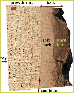
As you would expect, given the original publication, the images are excellent. The navigation of the site fits the definition of user-friendly. There are pages "Macroscopic characteristics" and "Microscopic characteristics" that provide an introduction to terminology, with an illustrated glossary and comments on the diagnostic value of different characteristics. There are multiple ways to get to species descriptions with 1) multiple images, particularly of transverse sections, to illustrate within species variation, 2) brief descriptions of the features visible in each plane of view, and 3) comments on diagnostic features, including comments such as "The wood of the three Quercus species (Q. robur, Q. petraea, Q. pubescens) cannot be differentiated on the basis of their wood anatomy."
Thumbnails on the species pages are "live" and provide access to larger images. "Species list" is alphabetical by genus and species list of all species (about 130), "Coniferous wood" and "Dicotyledon wood" list species for those groups.
The identification key is a synoptic key, with characteristics presented in tabular form. From the key you can access definitions and images for the key characters. It took me a bit time to realize that "Wood types" at the top of the key was a pull-down menu to take you to the major subdivisions within the table: Coniferous wood, Ring-porous wood, Diffuse- and semi-ring-porous wood, Diffuse- and semi-ring-porous wood with aggregate rays.
This site is valuable, as was the 1990 book, for showing both the variability of wood as well as documenting what many find an unpleasant fact, namely that many tree species share similar wood anatomy. For any one who teaches wood anatomy, this is a most useful resource. The site is a model for attractiveness and usability.
Identification key
Coniferous wood | Wood without vessels (pores), with tracheids only | |||||
| Pitting in rays | Resin canals | Transversal tracheids | Spiral thickenings in tracheid walls | Species | Key characteristics | |
| large (pinoid pits) | present | present | absent | Pinus sylvestris | Transversal tracheids with dentated walls, cannot be differentiated from P. mugo and P. nigra. | |
| Pinus mugo | Transversal tracheids with dentated walls, cannot be differentiated from P. sylvestris and P. nigra. | |||||
| Pinus nigra | Transversal tracheids with dentated walls, cannot be differentiated from P. sylvestris and P. mugo, sometimes early-/latewood transition seems more abrupt than in the two other species. | |||||
| Pinus cembra | Transversal tracheids with smooth walls, in general small growth rings, latewood zones always narrow. | |||||
| Pinus strobus | Transversal tracheids with smooth walls, in general larger growth rings, latewood zones narrow, similar to P. cembra. | |||||
| small | present | present | absent | Picea abies | Transition from early- to latewood continuous. Resin canals with thick-walled epithelial cells. Bordered pits in axial tracheids generally uniseriate. | |
| Larix decidua | Transition from early- to latewood abrupt. Resin canals with thick-walled epithelial cells. Bordered pits in axial tracheids often biseriate. | |||||
| present | Pseudotsuga taxifolia | Transition from early- to latewood abrupt. Resin canals with thick-walled epithelial cells. Fine spiral thickenings. | ||||
| absent | absent | absent | Abies alba | Transition from early- to latewood abrupt. In radial section the tangential ray cell walls show distinct nodular chains. | ||
| Juniperus communis | Colored deposits in parenchyma cells, smooth walled ray cells. | |||||
| present | Taxus baccata | Distinct spiral thickenings. | ||||
Dicotyledon wood | Wood with vessels (pores) | ||||
Ring-porous wood | |||||
| Ray width | Perforation plates | Spiral thickenings | Species | Key characteristics | |
| uniseriate | simple | absent | Castanea | Latewood with dendritic pore arrangement, rarely biseriate rays. | |
| bi- to triseriate | simple | absent | Fraxinus | Latewood pores solitary or in small radial groups, thickwalled. | |
| present | Hippophaë | Ring-porous, sometimes semi-ring-porous, rays generally storied. | |||
| 3- to 5-seriate | simple | present | Ulmus | Pores, vascular tracheids and parenchyma in latewood in tangential to slightly oblique bands. | |
| Robinia | Pores, vascular tracheids and parenchyma in latewood in clusters to short bands, conspicuous tyloses. | ||||
| >5-seriate | simple | absent | Vitis | In the narrow latewood pores in radial files and small groups. Rays very wide. Vessels with scalariform pits. | |
| present | Clematis | Latewood generally narrow, growth ring boundaries festoonlike, large rays. | |||
| Berberis | Ring-porous to semi-ring-porous, latewood pores and vascular tracheids in clusters with a tangential to diagonal or dentritic orientation. | ||||
| Laburnum | Growth ring boundaries festoonlike, rays often over 5 cells wide, gum deposits in the heartwood vessels. | ||||
| Ailanthus | Often slightly oblique to tangential parenchyma bands, often indistinct ring-porous, variable. | ||||
| uni- and multiseriate | simple | absent | Quercus | Dendritic pore groups in the latewood, uniseriate and very broad rays. | |
| present | Rosa | Broad rays often over 10 mm high. | |||
Diffuse- and semi-ring-porous wood | ||||||
| (uniform distribution of pores) | ||||||
| Ray width | Ray type | Perforation plates | Spiral thickenings | Species | Key characteristics | |
| uniseriate | homo- geneous | simple | absent | Populus | Large and simple ray-vessel pits. | |
| present | Aesculus | Pores solitary or in radial rows of two to some pores. | ||||
| Euonymus | Numerous small pores. | |||||
| scalariform | absent | Alnus viridis | Pores in radial multiples, rarely in clusters, scattered. | |||
| Alnus glutinosa Alnus incana Corylus | See aggregate rays. | |||||
| Betula nana Betula humilis | Extremely small numerous ray-vessel pits, occasionally bi- to 4-seriate rays. | |||||
| hetero- geneous | simple | absent | Salix | Large and simple ray-vessel pits. | ||
| present | Daphne | Pores loosely packed in dendritic patterns. | ||||
| Ray width | Ray type | Perforation plates | Spiral thickenings | Species | Key characteristics | |
| bi- to tri- seriate | homo- geneous | simple | absent | Juglans | Pores large, infrequent, solitary or in radial rows of 2 to 4 cells. | |
| absent to (sometimes: occasionally) present | Maloideae: Amelanchier, Cotoneaster, Crataegus, Cydonia, Mespilus, Pirus, Sorbus | Numerous to very numerous small pores, often indistinctly semi-ring-porous, occasionally with fine spiral thickenings. | ||||
| present | Hippophaë | Generally semi-ring-porous, sometimes ring-porous, rays generally storied. | ||||
| Acer | Pores widely spaced, solitary or in radial files of 2 to 3 pores. | |||||
| Prunus | See 3- to 5-seriate rays, slightly heterogeneous. | |||||
| Tilia | Vessel outlines angular, radially orientated pore files and clusters. Rays flare along growth ring boundaries. | |||||
| scalariform | absent | Betula | Extremely small numerous ray-vessel pits, scattered pores in radial files of 2 to 4, or clusters. | |||
| hetero- geneous | simple | present | Frangula | Generally semi-ring-porous, pores rather widely spaced, especially in latewood. | ||
| Lonicera | Multiseriate rays conspicuously heterogeneous with numerous rows of square and upright marginal cells. | |||||
| Ligustrum | Rays often with 1-2 (4) rows of square and upright marginal cells. | |||||
| Ostrya | Pores infrequently, in radial multiples of 2 to 10 pores. | |||||
| Prunus | See 3- to 5-seriate rays, slightly heterogeneous. | |||||
| Sambucus | See 3- to 5-seriate rays. | |||||
| scalariform | absent | Buxus | Narrow pores, round to oval scalariform perforation plates with mostly <10 bars. | |||
| Viburnum | Rays conspicuously heterogeneous, perforation plates scalariform with >20 bars. | |||||
| Cornus | Rays markedly heterogeneous, more likely 3- to 5-seriate, scalariform perforation plates with many bars. | |||||
| present | Viburnum | Rays conspicuously heterogeneous, perforation plates scalariform with >20 bars. Fine spiral thickenings in the fibre tracheids: Viburnum lantana. | ||||
| Ray width | Ray type | Perforation plates | Spiral thickenings | Species | Key characteristics | |
| 3- to 5- seriate | homo- geneous | simple | absent | Juglans | Pores large, infrequent, solitary or in radial rows of 2-4 cells. | |
| present | Prunus | Diffuse-porous to semi-ring-porous, pores solitary or in radial rows of two to some cells or in clusters, gum deposits in heartwood vessels. Rays in some species frequent >5-seriate. | ||||
| Acer | Pores widely spaced, solitary or in radial files of 2 to 3 pores. | |||||
| Tilia | Vessel outlines angular, radially orientated pore files and clusters. Rays flare along growth ring boundaries. | |||||
| scalariform | absent | Betula | Extremely small numerous ray-vessel pits, scattered pores in radial files of 2 to 4, or clusters. | |||
| hetero- geneous | simple | absent | Sambucus | Pores in clusters, mostly marginal bands of thin-walled vascular tracheids. Rays with sheath cells. | ||
| present | Prunus | Diffuse-porous to semi-ring-porous (P. armeniaca to ring-porous), pores solitary or in radial rows of two to some cells or in clusters, gum deposits in heartwood vessels. Rays in some species frequent >5-seriate. | ||||
| scalariform | absent | Cornus | Rays markedly heterogeneous, 3- to 5-seriate, scalariform perforation plates with many bars. | |||
| present | Ilex | Pores small, in long radial files, rays often up to 4mm high. | ||||
| Ray width | Ray type | Perforation plates | Spiral thickenings | Species | Key characteristics | |
| > 5-seriate often or predomi- nantly | homo- geneous | simple | absent | Fagus | Diffuse- to semi-ring-porous, rays very large, perforation plates both simple and scalariform. | |
| Platanus | Similar to Fagus, more often large rays, pits in vessel walls in horizontal rows. | |||||
| Hedera | Pores in clusters, predominantly tangentially orientated. | |||||
| present | Clematis | Very large pores, see ring-porous wood. | ||||
| Berberis | See ring-porous wood. | |||||
| scalariform | absent | Fagus | Diffuse- to semi-ring-porous, rays very large, perforation plates both simple and scalariform. | |||
| Platanus | Similar to Fagus, more often large rays, vessel pits opposite, in horizontal rows. | |||||
| hetero- geneous | simple | absent | Hedera | Pores in clusters, predominantly tangentially orientated. | ||
| Vitis | See ring-porous wood. | |||||
| scalariform | present | Ilex | Pores small, in long radial files, rays often up to 4mm high. | |||
| Ray width | Ray type | Perforation plates | Spiral thickenings | Species | Key characteristics | |
| uniseriate and/to multiseriate | homo- geneous to hetero- geneous | simple | absent | Fagus | Diffuse- to semi-ring-porous, rays very large, perforation plates both simple and scalariform. | |
| ClematisQuercus Fraxinus | Ring-porous wood with very narrow rings appears sometimes similar to diffuse-porous wood, see ring-porous wood. | |||||
| present | Rosa | Ring-porous wood with very narrow rings appears sometimes similar to diffuse-porous wood, see ring-porous wood. | ||||
| Prunus | Diffuse-porous to semi-ring-porous (P. armeniaca to ring-porous), pores solitary or in radial rows of two to some cells or in clusters, gum deposits in heartwood vessels. | |||||
| scalariform | absent | Ribes | Rays often with sheath cells. | |||
| present | Fagus | Diffuse- to semi-ring-porous, rays very large, perforation plates both simple and scalariform. | ||||
Diffuse- and semi-ring-porous wood with aggregate rays | ||||||
| (uniform distribution of pores) | ||||||
| Ray width | Ray type | Perforation plates | Spiral thickenings | Species | Key characteristics | |
| uniseriate and irregularly multiseriate | homo- geneous to hetero- geneous | simple | present | Carpinus | Pores in long radial files. | |
| scalariform | absent | Alnus glutinosa | Pores in radial multiples, perforation plates with generally more than 10 bars. | |||
| Alnus incana | Cannot be differentiated from A. glutinosa. | |||||
| Corylus | Pores in radial multiples, often in dendritic arrangement, perforation plates with 5 to 10 bars. | |||||
| present | Corylus | Fine spiral thickenings are frequent. | ||||
Diffuse- and semi-ring-porous wood, dendritic groups of pores | ||||||
| Ray width | Ray type | Perforation plates | Spiral thickenings | Species | Key characteristics | |
| 2- to 3- seriate | homo- geneous to hetero- geneous | simple | present | Rhamnus | Dendritic pore distribution. | |
| Daphne | Pores loosely packed in a dendritic pattern. | |||||
Diffuse- and semi-ring-porous wood, pores hardly differentiable from axial and ray parenchyma | ||||||
| Species | Key characteristics | |||||
| Viscum album | Vessels, parenchyma and growth ring boundaries indistinct. | |||||
Macroscopic characteristics |
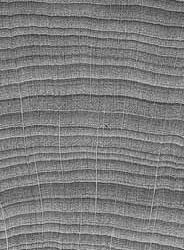
| Recent wood can often be identified by macroscopic characteristics, particularly by colour, gloss, odour, weight and structure. As such characteristics are generally modified or destroyed in fossil, historic or carbonized wood, only a few species or species groups of the indigenous flora can be identified with the naked eye or only with the aid of a magnifier (5 to 20x). | |||||
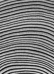 |  |  | |||
Coniferous woodIn coniferous wood it is possible to distinguish the species which have resin canals from those which do not. The transition from earlywood to latewood can be sharp or continuous. |
Ring porous
The diameter of the pores in the earlywood is much greater than the diameter of the pores in latewood. |
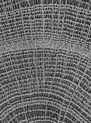
Semi-ring to diffuse porous
In semi-ring porous woods, the pores are more numerous in earlywood, in diffuse porous woods the size of pores and distribution is more regularely. Arrangement and size of pores, fine and large rays differ from species to species. | |||
| An identification key based on macroscopis characters is not given here as it exits e.g. In Bosshard (1974/75) or Gottwald (1958). | |||
 |  |  | |
Preparation of wood for microscopic examination | Recent, healthy wood | Secondary wood modifications |
Glossary & Links |
Glossary | |
| English and French glossary of wood (Faculté de foresterie et de géomatique, Université Laval, Québec, Canada) sylva.sbf.ulaval.ca/foret/glossAnat/index.html | |
| English glossary (School of Biological Sciences, The University of Sydney) BUGS.bio.usyd.edu.au/2003A+Pmodules/glossary.html | |
| English glossary (Biological Sciences Center,University of Rhode Island) www.uri.edu/cels/bio/plant_anatomy/glossary.html | |
| Multilingual glossary of dendrochronology (WSL, Switzerland) www.wsl.ch/dbdendro/glossary/index_EN | |
Links | |
| Forest Products Laboratory www2.fpl.fs.fed.us/ | |
| IAWA International Association of Wood Anatomists www.iawa-website.org |
We appreciate your feedback! |
| Any comments, concerns or compliments will help us to improve the wood anatomy website. Please send your feedback! |
http://www.woodanatomy.ch/review.html


